How In Vivo Infection Models Generate Actionable Evidence Beyond Traditional Antimicrobial Claims
Medical device innovation doesn’t stop performance, usability, or even traditional biocompatibility. For many products, the real-world risk that matters most shows up after the device enters the clinical environment: microbial contamination, colonization, biofilm formation, and infection. These issues can influence patient outcomes, hospital workflows, and a manufacturer’s ability to support product claims and adoption.
That’s where microbial animal model testing, often referred to as in vivo infection models or antimicrobial efficacy studies, can play a pivotal role. While these studies may involve implantation or surgical procedures (which can make them look like “biocompatibility studies” at first glance), they are equally grounded in microbiology: selecting organisms, preparing and verifying inocula, designing recovery methods, quantifying colony forming units (CFUs), and interpreting results in the context of host immunity and clinical use.
Importantly, these studies do not replace or alter sterility assurance. Sterility assurance remains focused on validated sterilization processes and sterility testing. Instead, microbial animal model testing evaluates how a device behaves after use, particularly when microbial contamination is clinically relevant. This distinction is essential for manufacturers developing devices intended to reduce infection risk, improve handling, or support antimicrobial or anti-infective claims.
What is Microbial Animal Model Testing?
Microbial animal model testing evaluates medical devices or materials in a living system under a controlled microbial challenge. The goal is to understand how bacteria interact with a device in a biologically relevant environment, including tissue response, host immunity, and procedural variables that cannot be fully replicated in vitro.
These studies commonly assess:
- Microbial colonization and recovery from devices or surrounding tissue
- Clinical signs of infection at surgical or implantation sites
- Quantitative microbial burden (e.g., colony forming units, CFUs)
- Tissue response and wound healing through histopathology
- Comparative performance between control and test articles
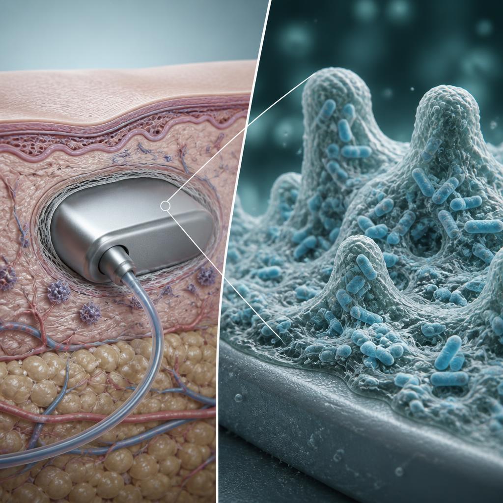
A representative example is an in vivo sutured wound model in rats developed to evaluate bacterial colonization following inoculation with Staphylococcus aureus. In this model, sutures were placed in muscle incisions, inoculated with defined bacterial concentrations, and evaluated for infection, bacterial recovery, and tissue response. The study demonstrated how dosing conditions and recovery methods directly influence the ability to detect meaningful microbial outcomes.
A representative example is an in vivo sutured wound model in rats developed to evaluate bacterial colonization following inoculation with Staphylococcus aureus. In this model, sutures were placed in muscle incisions, inoculated with defined bacterial concentrations, and evaluated for infection, bacterial recovery, and tissue response. The study demonstrated how dosing conditions and recovery methods directly influence the ability to detect meaningful microbial outcomes.
This type of work highlights why microbial animal model testing is both microbiology-driven and biologically complex—success depends as much on microbial recovery strategy and inoculum verification as it does on surgical technique.
Why “Antimicrobial Efficacy” is Not the Whole Story
These studies are often labeled as “antimicrobial efficacy studies,” but that term can be limiting. While many projects involve antimicrobial coatings or materials, not all microbial animal model studies include an antimicrobial agent.
In practice, these models are equally valuable for evaluating:
- Device designs that may influence infection risk through handling or implantation technique
- Surface treatments that alter bacterial attachment without releasing an antimicrobial
- Materials intended to reduce microbial transfer or biofilm formation
- Procedural workflows that may introduce contamination during device placement
For example, microbial animal models have been used to investigate whether device handling during surgery contributed to reported infection rates. In such cases, study outcomes supported design modifications that reduced handling complexity and improved implantability—demonstrating infection risk reduction through design, not chemistry.
This broader perspective allows manufacturers to evaluate infection-related performance without restricting the study to antimicrobial claims alone.
When a Manufacturer Should Consider Microbial Animal Model Testing
Microbial animal model testing is often considered when manufacturers need evidence that goes beyond benchtop microbiology, particularly when clinical use introduces variables that are hard to replicate in vitro. Common drivers include:
Supporting Product Claims
When a device is intended to reduce infection risk or demonstrate antimicrobial benefit, in vivo data can provide biologically relevant evidence to support those claims—especially when clinical simulation is important for market acceptance.
Evaluating Biofilm-Related Risk
For indwelling or implanted devices, biofilm formation presents a persistent challenge. Animal models allow assessment of bacterial persistence and colonization in the presence of host immune response.
Understanding Infection Pathways Linked to Use and Handling
Design features, implantation time, surface exposure, and procedural complexity can all influence contamination risk. Microbial animal models allow these factors to be evaluated under controlled conditions.
Device Types Suited for Microbial Animal Model Testing
Microbial animal models have been applied across a wide range of medical devices, including:
- Sutures with antimicrobial coatings
- Orthopedic implants, where post-operative infection is a major clinical concern
- Catheters / PIC lines / urinary catheters, where bacterial migration and biofilm are common themes
- Wound care products (bandages, dressings, hydrogels), with or without antimicrobials
- Surgical drapes and incision-associated products designed to reduce bacterial transfer from skin
- Skin-applied barrier materials (e.g., cyanoacrylate “glues”) intended to immobilize skin bacteria and reduce incision contamination
- Meshes/tissue reinforcement products, where multiple organisms may be requested to reflect risk profiles

The common factor is clinical exposure to microorganisms, not the presence of an antimicrobial ingredient.
How Microbial Animal Studies Are Designed
Microbial animal model studies are closer to engineered systems—because you’re balancing two living variables: the microorganism and the host. Results can be variable, and establishing a dose that persists and creates a consistent infection/colonization signal can be challenging. Key design elements often include:
1) Selecting the animal model
Species selection depends on multiple factors, including:
- Size and geometry of the device/implant (what can be realistically implanted and assessed)
- Intended clinical site (e.g., bone-related applications may drive toward models that accommodate orthopedic placement)
- Precedent in literature or prior programs, especially when trying to replicate an established model
- Cost and feasibility, where starting in a smaller model may allow greater sample sizes and iterative learning
Most microbial animal model studies use healthy animals. However, models with suppressed immune response, such as neutropenia-induced rodents, can be used when prolonged bacterial persistence is required. These models are particularly useful when evaluating devices intended for use in patient populations with compromised immunity.
2) Choosing microorganisms (and sometimes cocktails)
Organisms are selected based on clinical relevance, often including Gram-positive and Gram-negative bacteria associated with healthcare-acquired infections. In some cases, multiple organisms are evaluated, either individually or as part of a cocktail, with careful consideration of microbial competition and study complexity.
3) Establishing inoculum preparation and verification
Accurate preparation and confirmation of microbial dose are critical. Plate counts and verification steps ensure that the challenge level is known and reproducible.
4) Planning recovery methods (a crucial early milestone)
Before definitive studies begin, recovery methods are established and validated to ensure microorganisms can be reliably recovered from the device or tissue. This step is essential for meaningful quantitative results.
5) Building in pilot phases and iteration
Due to biological variability, pilot phases are often necessary to refine dose, exposure time, recovery methods, and procedural variables. Iteration improves consistency and reduces uncertainty before scaling to larger studies.
6) Determining endpoints and analysis plans
Typical endpoints include CFU recovery, gross observations, histopathology, and statistical evaluation. Robust study design and appropriate animal numbers are essential to support meaningful interpretation.
What Manufacturers Can Gain from Microbial Animal Model Testing
When designed well, these studies can generate evidence that supports decisions across R&D and commercialization:
Risk reduction: Understand microbial performance in a living system where immune response and biological complexity affect outcomes
Early screening: Compare materials, coatings, or surface treatments before committing to a definitive path
Design optimization: Identify procedural or handling-related risks that may be mitigated through design changes
Claim support: Build a stronger evidence package when in vivo relevance is needed to back messaging
Frequently Asked Questions (FAQs)
Does microbial animal model testing replace sterility assurance testing?
No. Sterility assurance focuses on validating sterilization processes and demonstrating sterility. Microbial animal model testing evaluates how a device behaves in vivo under microbial challenge conditions and can inform infection-related performance—but it does not replace sterility assurance.
Do these studies always require an antimicrobial coating or agent?
Not necessarily. While many studies involve antimicrobial coatings (e.g., antimicrobial sutures), projects may focus on handling, design, or surface effects that influence infection outcomes without any antimicrobial ingredient.
Are there standardized ISO or ASTM in vivo guidelines for these studies?
There is no single ISO or ASTM standard governing in vivo antimicrobial or microbial challenge testing. These studies are typically custom designed based on the device and intended use.
Which animal species are commonly used?
It depends on the device and clinical use case. Rats, mice, guinea pigs, and rabbits are used most, with larger species, such as pigs, used less frequently. Species selection is based on multiple factors such as implant size, intended anatomical location, precedent in literature, and feasibility.
Why do these studies often require pilots or iteration?
Biological systems introduce variability. Pilot studies allow refinement of microbial dose, recovery methods, and procedural parameters to improve consistency and data quality.